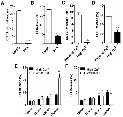
Figure 3. Suppression of Cx31R42P induced cell death by connexin hemichannel blocker and high extracellular calcium.
(A) Quantification of Cx31R42P cells with SN after treatment with 200 µM FFA or control solvent DMSO. (B) The rate of LDH release of Cx31R42P cells after treatment with 200 µM FFA or control solvent DMSO. (C) Quantification of Cx31R42P cells with SN incubated at 26°C with physical or high Ca2+ o. (D) The rate of LDH release of Cx31R42P cells incubated at 26°C with physical or high Ca2+ o. (E–F) The rate of LDH release of Cx31R42P cells (E) and Cx31WT cells (F) at different time points after Ca2+ o treatment was washed out. Error bars represent SEM. Two stars: P<0.01; three stars: P<0.001.

