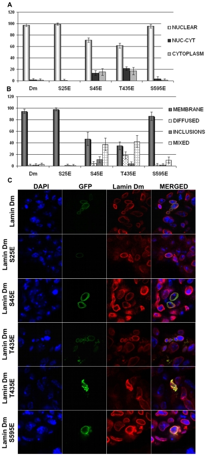
Figure 4. All lamin Dm mutants localize to nuclear lamina in transfected Drosophila S2 cells but lamin Dm S45E and T435E mutants show significantly different distribution.
Localization of fusion GFP-lamin Dm and mutant proteins after 48 h (C) post-transfection into Drosophila S2 cells visualized under a confocal microscope and quantitative analyses of appearance of the particular phenotypes (A and B). Cells were stained for DNA with DAPI, for endogenous lamin Dm with mouse monoclonal antibodies ADL67. Exogenous lamin Dm proteins were visualized by eGFP fluorescence. Panel A demonstrates statistical analyses of distribution of lamin fusion proteins in nucleus only in nucleus and cytoplasm and in cytoplasm only. Panel B demonstrates statistical analyses of fusion proteins' localization to nuclear envelope and nuclear lamina (membrane), diffused phenotype, inclusion bodies phenotype and mixed phenotype respectively. Single confocal sections through the center of nuclei are shown. 200 cells were analyzed.

