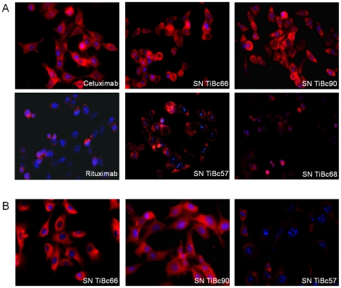
Figure 3. Representative immunofluorescence images of tumor cells showing positive binding of both membranous as well as intracellular target structures by TiBc-derived IgGs.
(A) HCT116 and (B) HROC46 tumor cells were stained with TiBc-derived IgGs and Alexa 546-conjugated anti-human IgG. Cell nuclei were counterstained with DAPI. Therapeutic antibodies Cetuximab and Rituximab served as positive and negative control, respectively. Original magnification ×40.

