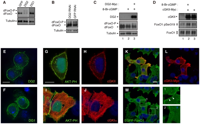
Figure 3. DG2 modulates FoxO in vivo.
(A) dFoxO and the indicated transgenes were expressed in the Drosophila eyes using the GMR-GAL4 driver. Extracts from brain tissues were subjected to western blot analysis. dFoxO-P; a phosphorylated form of dFoxO. (B) DG2 RNAi or GFP RNAi constructs were expressed in the Drosophila brain using the elav-GAL4 driver. Western blot analysis for endogenous dFoxO was carried out as in (A). (C) Drosophila S2 cells were transfected with or without C-terminally Myc-tagged DG2 (DG2-Myc). Thirty-six hrs post transfection, cells were treated with or without 10 µM 8-Br-cGMP for 30 min. Cell lysate were then subjected to western blot analysis. (D) Human 293T cells were transfected with or without cGKII-Myc, and were treated with 8-Br-cGMP as in (C). Phosphorylation of the S319 site in endogenous FoxO1 was detected with phospho-specific antibody. (E, F) S2 cells expressing DG2-Myc (E) or DG1-Myc (F) were visualized with anti-Myc antibody (green), by counterstaining with DAPI (blue color). (G–J) HeLa cells expressing AKT-PH-GFP (green) along with cGKII-Myc (G, H) or cGKI-Myc (I, J) were visualized with anti-Myc antibody (red), by counterstaining with DAPI (blue color). AKT-PH-GFP was used for a marker protein of the plasma membrane [62]. (K–M) Flp-In T-REx-293 cells harboring EGFP-FoxO1 gene were transiently transfected with cGKII-Myc, and EGFP-FoxO1 was induced with doxycycline. Enlarged views of the plasma membrane regions in cGKII-positive (Box1) and negative (Box2) cells are also shown in (M). Accumulation of FoxO1 along with cGKII in the plasma membrane is indicated by arrowheads. Scale bars = 5 µm for (E, F), 25 µm for (G–J) and 10 µm for (K–M).

