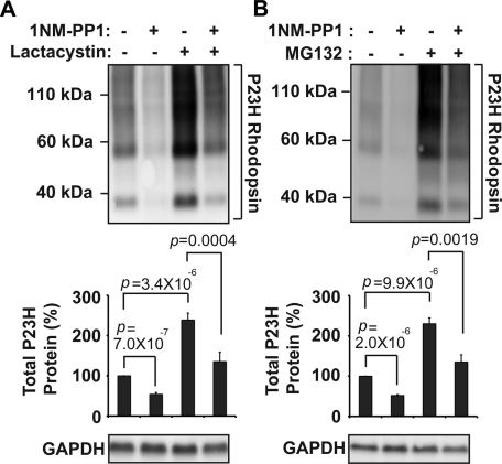
FIGURE 4:
P23H rhodopsin protein degradation depends on proteasome function. (A) P23H rhodopsin was expressed in cells bearing IRE1[I642G], and lactacystin (1 μM) and/or 1NM-PP1 (5 μM) was added for 24 h as indicated. Rhodopsin protein levels were detected by immunoblotting and quantified. (B) P23H rhodopsin was expressed in cells bearing IRE1[I642G]. MG132 (1 μM) and/or 1NM-PP1 (5 μM) were applied for 24 h as indicated. (A, B) GAPDH protein levels were assessed as a protein loading control. Immunoblots are representative of three independent experiments. Error bars represent SDs from three experiments. The p values were determined by Student's t test analysis.

