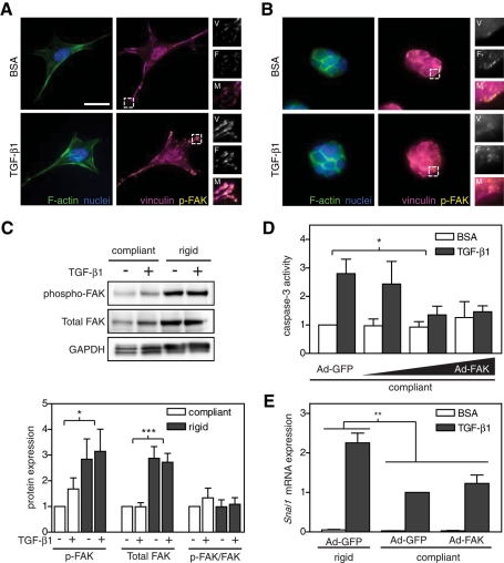
FIGURE 6:
Effect of matrix rigidity and TGF-β1 on the actin cytoskeleton and focal adhesion formation in NMuMG cells. (A,B) Immunofluorescence images of F-actin (green), nuclei (blue), vinculin (magenta), and phospho-FAK (yellow) on rigid (8 kPa) (A) and compliant (0.4 kPa) (B) gels. Inset shows magnification of vinculin (V), phospho-FAK (F), and merged (M) images. (C) Western blot and quantification of phospho-FAK (125 kDa), total FAK (125 kDa), and GAPDH (38 kDa). Caspase-3 activity (D) and Snai1 mRNA expression (E) in NMuMG cells infected with Ad-GFP or Ad-FAK cultured on rigid and compliant gels treated with TGF-β1. n = 4 ± SEM. *p < 0.05; **p < 0.01; ***p < 0.001. Bars, 25 μm.

