FIGURE 7:
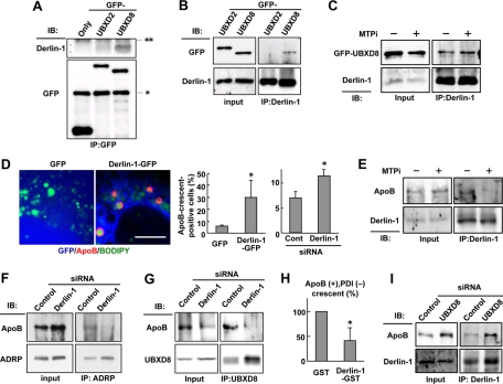
Inhibition of Derlin-1 function suppressed the cytoplasmic dislocation of ApoB. (A) Huh7 cells expressing GFP, GFP-UBXD2, or GFP-UBXD8 were lysed and immunoprecipitated by anti-GFP antibody. Endogenous Derlin-1 coprecipitated with GFP-UBXD8 significantly, whereas it bound with no GFP or little GFP-UBXD2. The IgG heavy and light chains are indicated by asterisk and double asterisks, respectively. (B) Huh7 cells expressing GFP-UBXD2 or GFP-UBXD8 were lysed and immunoprecipitated by anti–Derlin-1 antibody. GFP-UBXD8 coimmunoprecipitated significantly with endogenous Derlin-1, whereas little GFP-UBXD2 did so. (C) Huh7 cells treated with 10 μM ALLN alone or with 10 μM ALLN and 100 nM BAY13-9952 for 12 h were lysed, and endogenous Derlin-1 was immunoprecipitated. Comparable amounts of GFP-UBXD8 coprecipitated with Derlin-1 irrespective of the MTPi treatment. (D) Huh7 cells expressing GFP or dominant-negative Derlin-1 (Derlin-1–GFP; green) were labeled for ApoB (red) and LDs (blue). Bar, 10 μm. The frequency of ApoB-crescent–positive cells was significantly higher in cells expressing Derlin-1–GFP than in those expressing GFP. ApoB-crescents also increased by Derlin-1 knockdown. The average of results from three independent experiments is shown (Student's t test; *p < 0.05). (E) Huh7 cells were treated as in (C). ApoB coprecipitating with Derlin-1 was reduced significantly by the MTPi treatment for 12 h. (F) Huh7 cells transfected with control or Derlin-1 siRNA were incubated with 10 μM ALLN for 12 h. ApoB cross-linkable to ADRP with 1 mM DSP was reduced by Derlin-1 knockdown. (G) Huh7 cells were treated as in (F). ApoB coimmunoprecipitating with UBXD8 was reduced by Derlin-1 knockdown. (H) The dislocation assay by immunofluorescence microscopy. Huh7 cells were transfected with cDNA of either GST alone or Derlin-1–GST, treated with 10 μM ALLN for 12 h, and labeled for ApoB (red), PDI (green), and GST (blue). Derlin-1–GST was used instead of Derlin-1–GFP to employ the same fluorophore combination for ApoB and PDI as in Figures 5C and 6C. The proportion of ApoB+, PDI− spheres was significantly lower in Derlin-1-GST–positive cells than that in the control, indicating that the dominant-negative Derlin-1 abrogated the cytoplasmic dislocation of ApoB. The average of three independent experiments is shown. (I) Huh7 cells were transfected with control or UBXD8 siRNA. UBXD8 knockdown did not influence the amount of ApoB coimmunoprecipitating with Derlin-1.

