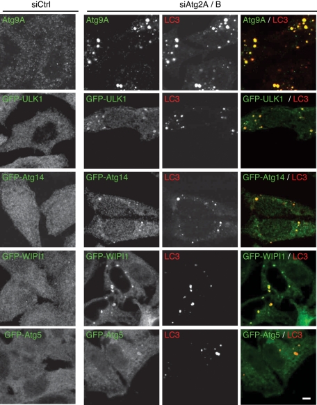
FIGURE 2:
Autophagy-related proteins accumulate in Atg2A/B-depleted cells. HeLa cells or HeLa cells stably expressing the indicated GFP-fused proteins were treated with control siRNA or siRNA against Atg2A and Atg2B. Cells cultured in regular medium were fixed and stained with the anti-Atg9A, anti-LC3 (CTB-LC3-2-1C; Cosmo Bio), and anti-GFP (A6455; Invitrogen) antibodies. Immunofluorescence images were obtained using a confocal microscope. Signal color is indicated by color of typeface. Scale bar, 5 μm.

