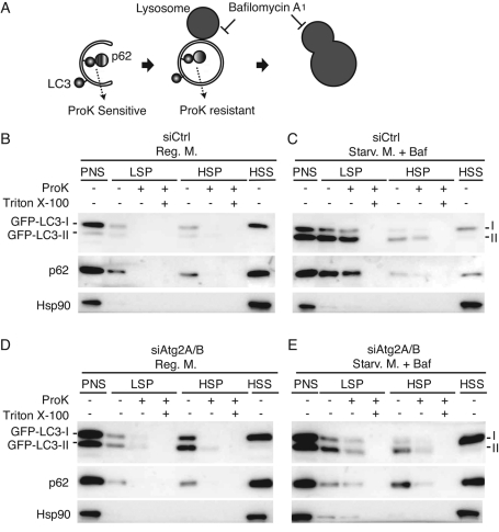
FIGURE 3:
Unsealed autophagosomes accumulate in Atg2A/B-depleted cells. (A) Schematic drawing of autophagosome formation and protease protection assay. (B–E) HeLa cells expressing GFP-LC3 were treated with control siRNA (B, C) or a mixture of siRNAs against Atg2A and Atg2B (D, E), and cultured in regular DMEM (B, D) or starvation medium containing 0.2 μM bafilomycin A1 (Baf) for 2 h (C, E). The PNS was separated into LSP, HSP, and HSS fractions and then analyzed by SDS–PAGE and immunoblotting using anti-GFP, anti-p62, and anti-Hsp90 antibodies. The subfractions were treated with proteinase K (ProK) with or without Triton X-100.

