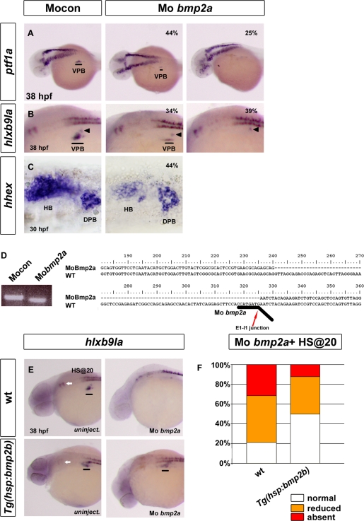
FIGURE 4:
Knockdown of bmp2a represses VPB specification. (A–C) Analysis of ventral pancreatic and hepatic markers in control morphants (left) and in bmp2a morphants (right) by in situ hybridization. (A) Lateral view of ptf1a expression at 36–38 hpf. (B) Laterial view of hlxb9la expression at 36–38 hpf. (C) Ventral view of hhex expression at 30 hpf. Percentage represents the proportion of morphants exhibiting a reduction (middle) or an absence (right) of gene expression. (D) Gel electrophoresis after RT-PCR (left) and sequencing result (right) illustrating the RNA splicing defects caused by the bmp2a morpholino in injected embryos. The sequence of the amplified bmp2a fragment was aligned with wt bmp2a cDNA and revealed a 86–base pair deletion in exon 1. Black line underlines the binding site of the bmp2a morpholino. Red arrow shows the junction between exon 1 and intron 1. (E, F) Lateral view of hlxb9la expression at 38 hpf in wt and Tg(hsp70l:bmp2b) in uninjected embryos (E) or in bmp2a morphants (F). Embryos were heat shocked at 20 hpf. (G) hlb9la expression in wt and Tg(hsp70l:bmp2b) embryos injected with MO bmp2a, heat shocked at 20 hpf, and fixed at 38 hpf. Data are presented as the percentage of embryos displaying normal (white), reduced (orange), or absent (red) expression of hlxb9la. Black lines indicate the VPB; black arrowheads indicate the dorsal pancreatic bud (DPB); white arrows indicate rhombomere 5 and 6 localization. HB, hepatic bud.

