FIGURE 5:
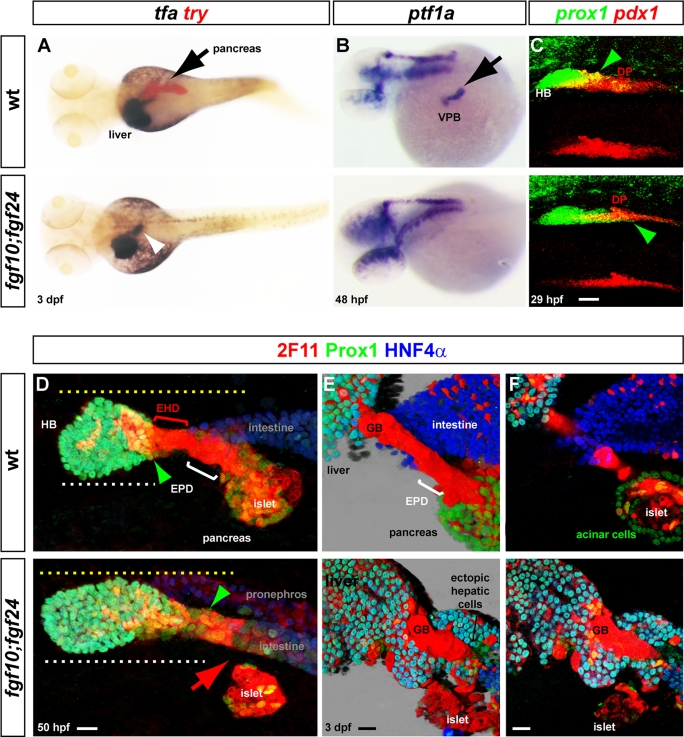
Absence of VPB cells and ectopic hepatic cells in fgf10; fgf24 compound mutants. (A) Dorsal view of transferrin (tfa) and trypsin (try) expression, comparing wt and fgf10; fgf24 mutants at 3 dpf. (B) Dorsolateral view of ptf1a expression comparing wt and double mutants at 48 hpf. (C) Confocal projection of the pdx1 and prox1 expression domains at 29 hpf showing the severe decrease of pdx1 in the anterior part of the pancreatic region and the extension of prox1 in fgf10; fgf24 mutants (green arrowheads). (D–F) Immunolabeling of the hepatopancreatic region with 2F11, Prox1, and HNF4α in wild-type and fgf10; fgf24 double mutants. (D) At 50 hpf, the ventral pancreas and the EPD are absent (red arrow) in fgf10; fgf24 mutants (confocal projections). (E, F) Morphology and differentiation of the hepatopancreatic ducts connecting the pancreas and liver to the intestine in fgf10; fgf24 double mutants at 3 dpf. (E) Three-dimensional blend projection. (F). Z-planes). Black arrows indicate pancreas and VPB; white arrowhead indicates ectopic tfa expression. EHD, extrahepatic duct; EPD, extrapancreatic duct; GB, gall bladder; islet, endocrine islet. Scale bar, 20 μm.

