
FIGURE 5:
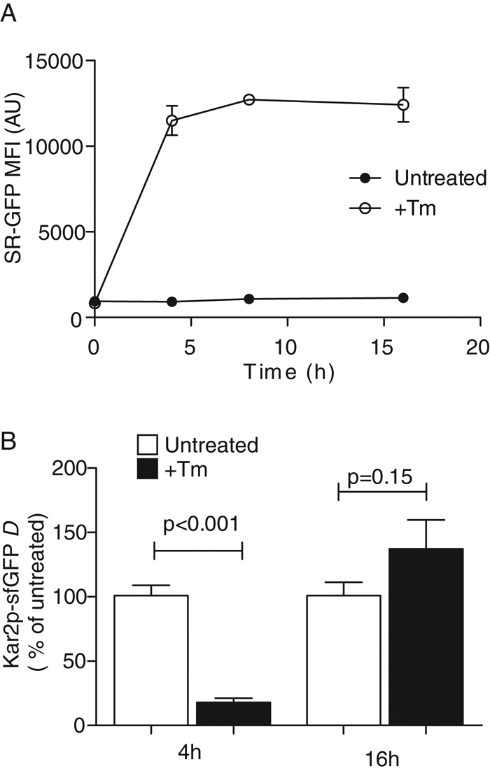
Kar2p availability reveals changes in the ER misfolded protein during adaptation. (A) Wild-type cells expressing the fluorescent splicing reporter (SR) consisting of GFP replacing the HAC1 open reading frame produce a fluorescence signal only when spliced by ire1. SR-GFP–expressing cells were treated with 1 μg/ml Tm, and GFP signal was measured over time using flow cytometry. (B) Mobility of Kar2p-sfGFP untreated cells or cells treated with 1 μg/ml Tm for 4 or 16 h was measured by FRAP. Data were normalized to the mean D values of untreated cells for both time points. At 4 h, Tm induces significant decrease in Kar2p-sfGFP mobility. Once stress is resolved and folding capacity of the ER is restored, the Kar2p-sfGFP mobility returned to the unstressed D values.