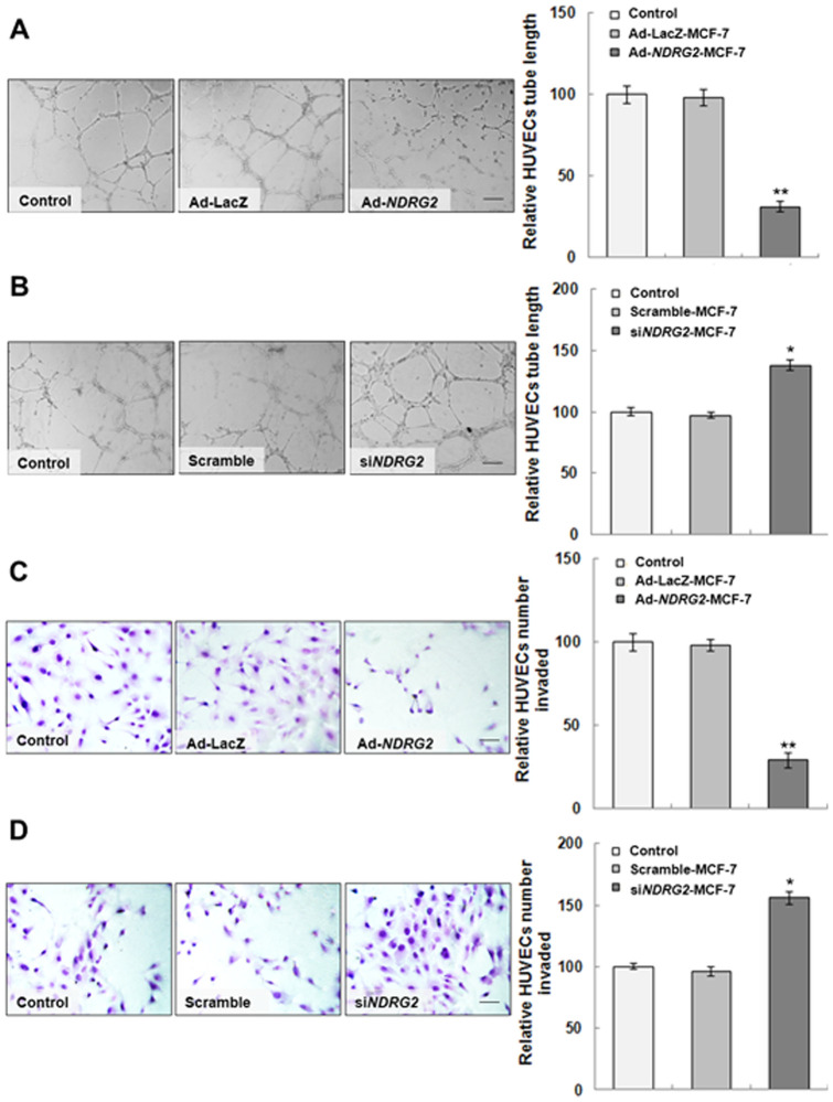
Figure 3. Effect of MCF-7 preconditioned media on HUVEC tube formation and invasion.
(A and B) The tube formation assay was performed as described in Materials and Methodss. HUVECs were plated on Matrigel and incubated under hypoxia for 6 h. Tube lengths were measured with Image-Pro Plus software; the histograms represent the quantification of the tube length of HUVECs. (C and D) The invasion assay was performed as described in Materials and Methods. After 4 h under hypoxia, the amount of invaded cells was calculated in five random high-power fields (400×), and the number from the control group was used as a control. The histogram represents the quantification of cells that invaded. The data are the mean ± standard deviation (SD) for three independent experiments. Statistical significance was assessed using one-way ANOVA and Student's t-test. * or ** indicates P<0.05 or P<0.01, respectively, when compared with the control. The Ad-NDRG2 panel of Figure 3A is excluded from this article's CC-BY license. See the accompanying retraction notice for more information.

