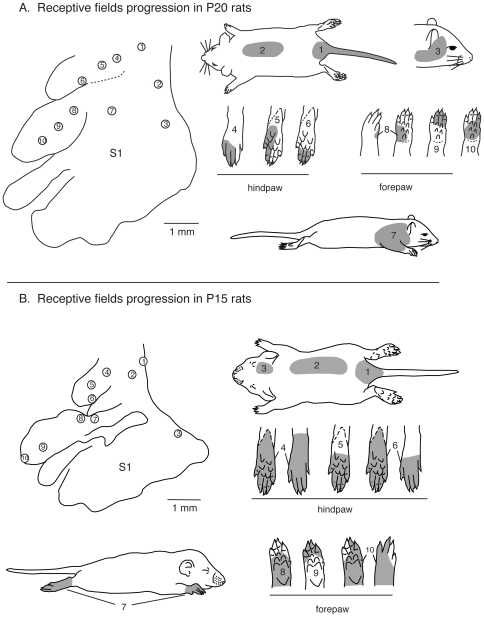
Figure 12. Receptive field progressions in P20 and P15 rats.
A) Progressions of recording sites in S1 in a P20 rat (left) and corresponding receptive fields for neurons at those sites (right). In P20 rats, the topographic organization is similar to that seen in adults. As recording sites progress from medial to lateral in the caudal portion of S1 (sites 1–3) corresponding receptive fields move from the tail, hindlimb and lower trunk to upper trunk and face. Compared to adults, receptive fields on the hindpaw (4–6) and forepaw (8–10) are larger and can encompass multiple digits, toes, or pads. B) Progressions of recording sites in S1 in a P15 rat (left) and corresponding receptive fields for neurons at those sites (right). In P15 rats the topographic representation is less well-organized and there is greater variability between animals. Receptive fields are larger and can encompass more than one body part (i.e., site 7). As recording sites progress from medial to lateral in the caudal portion of S1 (1–3) corresponding receptive fields move from the tail and lower trunk, to the middle trunk and head. Most often receptive fields are on the entire foot (4–5) or large portions of the forepaw (7–10). Compare this figure with the full map of the body illustrated in Figure 1 [102]. Conventions as in previous figures.

