FIG. 4.

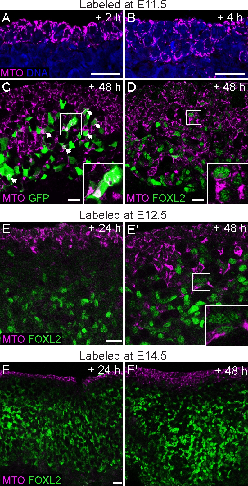
The surface epithelium is a source of new Sry-EGFP- and Foxl2-expressing cells in the fetal ovary. The ovarian surface was labeled with the cytoplasmic MitoTracker dye (MTO; magenta) at various stages of ovary development, and the samples were cultured for 2–72 h. A) Gonads from E11.5 embryos fixed after 2 h show that the dye is confined to the outermost cell layers. B) After 6 h, cell divisions in the coelomic epithelium generate labeled cells that move deeper inside the gonad. C) Ovary from an Sry-EGFP transgenic embryo labeled with MitoTracker at E11.5. Many EGFP-positive cells contained the label after 48 h of culture (overlap is white; arrowheads and inset). D) Similarly, in an E11.5 wild-type ovary cultured for 48 h, MitoTracker-labeled cells had ingressed to very deep layers, and many began to present nuclear FOXL2 staining (green; inset). E and E′) Gonads labeled at E12.5 displayed fewer ingressing cells that colabeled with FOXL2 after 24–48 h. F and F′) In samples labeled at E14.5, no ingression was observed after 24 h. Multiple cell layers were labeled after 48 h, but few ingressing cells were positive for FOXL2. Original magnification ×40 (A–E) or ×20 (F); bars = 20 μm.
