FIG. 6.
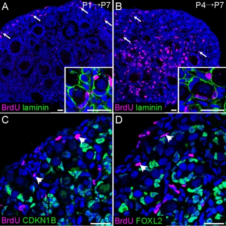
New granulosa cells arise in the ovarian cortex after birth. A and B) Ovaries of P7 pups exposed to BrdU at P1 (A) or P4 (B) and stained with antibodies against BrdU (magenta) and αlaminin (green) show BrdU-positive granulosa cells inside primordial follicles (arrows, insets). As expected, granulosa cells in actively dividing medullary follicles were heavily labeled in samples pulsed at P4, but not in samples pulsed at P1. We speculate that the label was titrated out during the week-long chase in the latter case. C and D) Somatic cells in and near the surface epithelium remain proliferative after birth. Ovaries from P1 mice were injected with BrdU 2 h prior to dissection and stained with antibodies against BrdU (magenta) and CDKN1B (C) or FOXL2 (D) (green). Arrowheads point to clusters of proliferative (CDKN1B-negative, BrdU-positive) FOXL2-negative somatic cells under the surface epithelium. Nuclei (blue) were stained with syto13. Original magnification ×20 (A and B) or ×40 (C and D); bars = 20 μm.

