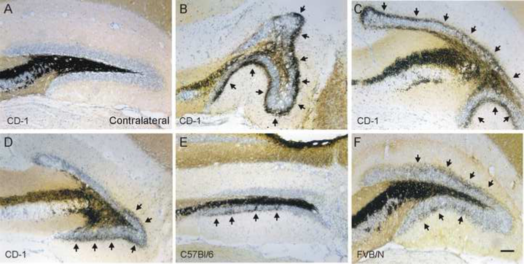
Figure 1.
MFS after cortical contusion injury is not uniform. Example Timm’s and Nissl stained sections of the dentate gyrus 8–12wk after CCI injury A. Representative image of hippocampus contralateral to the injury shows the absence of mossy fiber sprouting in the inner molecular layer (Timm score = 0). B–F. Representative images of Timm’s staining in the ipsilateral dentate gyrus near the injury epicenter. Note that the pattern of MFS and distortion of the granule cell layer is different in each section. MFS into the inner molecular layer is indicated by arrows. B–D. Sections obtained from CD-1 mice. Timm scores for these sections are B, 2; C, 3; D, 2. E. Section from a C57BL/6 mouse (Timm score = 1.5). F. Section from an FVB mouse (Timm score = 2). Scale bar is 100 μm.

