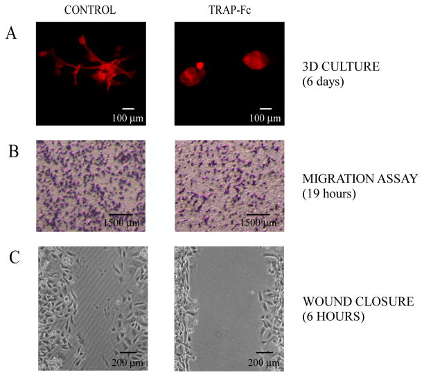
Figure 5. TRAP-Fc inhibits invasive growth of MDA-MB-231 mammary tumor cells in vitro.
(A) Eight-chambered plates were coated with an extracellular matrix (Matrigel™). MDA-MB-231 cells (2,000 cells/well) were mixed with medium containing Matrigel™ and then added to the chambers. Cells were incubated without or with TRAP-Fc (30 μg/ml), and phase contrast photomicrographs were captured six days later. (B) MDA-MB-231 cells (1.5×10⁵) were incubated in Transwell chambers in the absence or presence of TRAP-Fc (30 μg/ml). Cell migration across the filter separating the two compartments was measured 19 hours later and representative photographs of the lower faces of the filters were taken. Migration was normalized to the input number of cells. (C) MDA-MB-231 cells were plated on wound-healing inserts. Twenty-four hours later, plugs were removed, cells were treated with TRAP-Fc (100 μg/ml) and allowed to migrate. Snapshots captured after 6 hours are presented.

