Fig. (2).
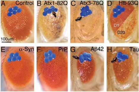
Disruption of the fly eye by expression of amyloidogenic proteins. (A-H) Microphotographs of the Drosophila compound eye. Higher magnifications show a detail from scanning electron microphotographs (artificially colored). (A) Control eye showing normal pigmentation and organization of the ommatidia. (B) Flies expressing Atx1-82Q show highly disorganized (glassy), depigmented eyes, with necrotic spots. High magnification shows poorly differentiated ommatidia with small bristles (arrow). (C) Flies expressing Atx3-78Q show disorganized and depigmented eyes with normal bristles. (D) Flies expressing Htt-93Q have normal eye structure at day 1 (left), but the underlying retina degenerates by day 20, resulting in patchy depigmentation (right). (E and F) Flies expressing 3 copies of wild type α-Syn (E) or wild type hamster PrP (F) have normal eye structure and pigmentation. (G and H) Flies expressing Aß42 (G) or wild type Tau (H) have small, disorganized, bumpy eyes resulting from fusion of ommatidia (arrows).

