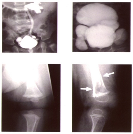
Fig. (8).
Connective tissue abnormalities in the patient with Menkes disease shown in Fig 7. Images on the left and right were taken just before treatment and at 2 years of age (also during the treatment period), respectively. Bladder diverticula formation (upper) and osteoporosis (lower) progressed despite treatment. Arrows show bone fractures.

