Figure 7.
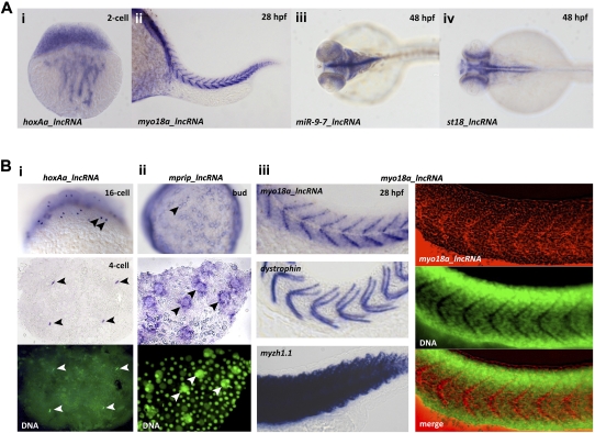
LncRNAs show tissue-specific and subcellularly restricted expression patterns. (A) Examples of lncRNAs with cell type–specific expression patterns at different stages of embryogenesis. Shown are in situ hybridization images with probes specific to the indicated lncRNAs. Expression is observed (i) in a two-cell stage embryo (cytoplasmic streaming from the yolk), (ii) in developing muscles, and (iii,iv) in distinct cells in the developing nervous system. (i,ii) Lateral views (anterior toward the left in ii); (iii,iv) dorsal views, anterior toward the left. (B) Examples of subcellularly localized lncRNAs. Bottom panels in i and ii (middle panel in iii, right) show a counterstain of the in situ image with the DNA-dye OliGreen (green). Black arrowheads point to subcellularly localized RNAs; white arrowheads point to the same position in the OliGreen-stained images. (i) Nuclear enrichment and association with chromatin (hoxAa-lncRNA); (top) 16-cell stage embryo with mitotically dividing nuclei; (middle, bottom) four-cell stage embryo. (ii) Enrichment at the nuclear periphery (mprip_lncRNA): (top) overview of a bud-stage embryo, showing accumulation of the lncRNA around nuclei of the yolk syncytial layer (YSL); (middle, bottom) close-up view of a dissected portion of the embryo shown in the top panel. Note that the lncRNA is specifically enriched around the large nuclei of the YSL but not around the small nuclei of the overlying cell-sheet. (iii) Enrichment at the myoseptum, the boundary between two adjacent myotubes (myo18a-lncRNA; top left, right); dystrophin mRNA (middle left) is a known marker of the myoseptum (Bassett 2003); myzh1.1 (myosin heavy chain) mRNA (bottom left) is detected throughout the somites (not subcellularly localized); and (right) myo18a-lncRNA (red, in situ) is enriched at the myoseptum, which is characterized by the absence of nuclei (regions of no green in the OliGreen-stained panel). Note that there is no overlap between red and green in the merge panel.

