
Fig. 2.
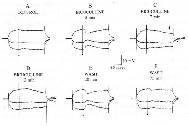
Bicuculline increased and then blocked the hyperpolarization evoked by fimbria stimulation. A: 3 responses to fimbria stimulation, triggered during hyperpolarizing or depolarizing current steps, are shown superimposed. Stimulation occurred at the dot. Membrane potential, −66 mV; resting potential, −82 mV. B: responses to the same fimbria stimulus are shown 5 min after 25 μM bicuculline was added to the buffer. Note the increase in IPSP amplitude. C: 7 min after bicuculline was added to the buffer, the IPSP was reduced and a second, later hyperpolarization was evident (arrow). D: at 12 min, the short latency IPSP was blocked, but the second hyperpolarization was not. E: 20 min after perfusion with drug-free buffer was resumed, stimulation produced a very large early IPSP but no late hyperpolarization. F: following prolonged perfusion with drug-free buffer, the response to fimbria stimulation was similar to control conditions.