Fig. 2.
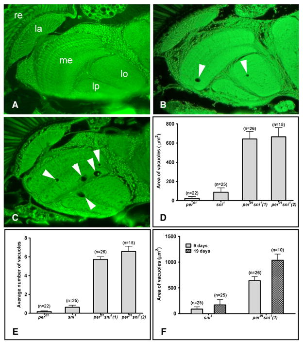
Interfering with the clock increases neurodegeneration in sni1 mutants. A–C) Paraffin head sections from 9 day-old males (scale bar=25 μm, re=retina, la=lamina, me=medulla, lo=lobula, lp=lobula plate). A) No vacuoles are detectable in the brain of a per01 fly. B) A sni1 fly brain shows a few vacuoles (arrows). C) Brains of per01 sni1 double mutant show increase in the size and number of vacuoles. D) Bar graph showing the mean±SEM area of all vacuoles/brain hemisphere. There is a significant difference between the sni1 and per01 sni1 line 1 (p=2.9×10⁻⁶), and sni1 and per01 sni1 line 2 (p=1.6×10⁻⁵). E) The mean number of vacuoles/brain hemisphere is increased in per01 sni1 compared to sni1 alone [sni1 to per01 sni1 (1): p=8.42×10⁻¹²; sni1 to per01 sni1 (2): p=6.5×10⁻⁸]. F) Comparison of the vacuolization between 9 and 19 day-old flies shows that the phenotype is progressive with age for both sni1 (p=0.036) and per01 sni1 line 1 (p=0.03). D–F) The number of brain hemispheres (n) examined to calculate the average values are indicated on the top of each bar.

