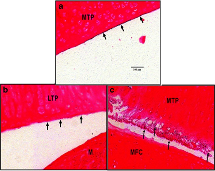
Fig. 3.
Micro-autoradiography analysis of knee joint sections after a single IA dose of [125I]LUB:1 to naïve rats or rats that underwent MT surgery. Naïve or MT rats were given a single 8.3 μg/knee dose of [125I]LUB:1, knees were collected at 24 and 168 h time points, and micro-autoradiography analysis followed by eosin staining of knee sections was conducted, as described in the text (study 4 in Table I, unilateral knee surgery). Representative digital images of the articular surface of the MTP in a naive animal a, LTP (without damage) of a MT animal b, and MTP (damaged) of the same MT animal c at 24 h time point. The intensity and extend of radioactive signals on the articular surfaces without damage a and b are significantly less than the damaged articular surface. Eosin staining; bar length in a equals 100 μm. Arrows point to the surfaces with radioactive signals. MT meniscal tear. MTP medial tibial plateau, LTP lateral tibial plateau, M meniscus, MFC medial femoral condyle

