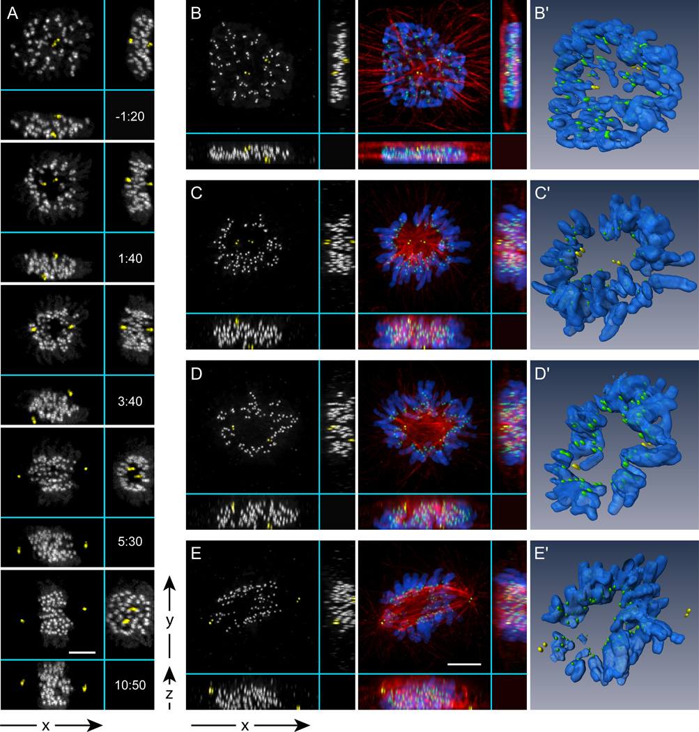
Figure 2. Multi-dimensional analysis of spindle assembly.
(A) Selected frames from a high-resolution 4-D time-lapse movie of a cell labeled with centrin1-GFP and CENP-A-GFP. For clarity, centrioles are pseudo-colored yellow. Notice that one centrosome is positioned above and the other – below the nucleus (V-cell). In less than 2 min after NEB a clear zone, void of chromosomes, develops between the separating centrosomes (1:40). As the spindle rotates, the zone persists as evident from the YZ view (5:30). Later, the chromosomes re-populate the central part of the spindle (10:50). Time shown relative to NEB in min:sec. (B–E) Immunofluorescence images and computer generated surface renderings (B'–E') of fixed RPE1 cells during early-to-mid prometaphase. The volume between the poles that is void of chromosomes is filled with high-density of microtubules (C–D; C'–D'). Once the spindle rotates to a vertical position a typical prometaphase morphology becomes apparent in the conventional XY view (E, E’). Bars, 5 µm.

