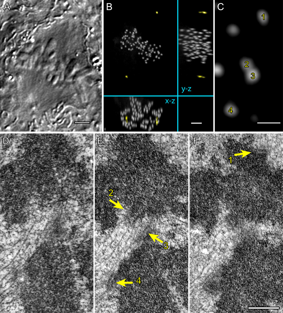
Figure 7. Fully congressed chromosomes can lack amphitelic attachment.
DIC image (A) and maximal-intensity XY, XZ, and YZ projections of GFP fluorescence (B) of a fixed metaphase RPE1 cell expressing centrin1-GFP and CENP-A-GFP. (C) A higher-magnification view (XY projection) showing two pairs (1–2 and 3–4) of sister chromosomes positioned within the metaphase plate. (D–F) Serial 70-nm thin sections through the area presented in (C) demonstrate that kinetochores 1, 2, and 4 are attached to microtubule in the end-on fashion, which implies that the chromosome in the top half of the image is amphitelic. In contrast, kinetochore 3 lacks end-on attachment and it is shielded from the top spindle pole by a mass of chromatin positioned in front of the kinetochore. This kinetochore laterally interacts with microtubules of the K-fiber that terminates within kinetochore 2. Bars in A and B, 5 µm. Bars in C–F, 0.5 µm.

