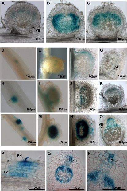
Figure 4.
Analysis of the tissue-specific expression pattern of WOX5 using promoter-reporter fusion in supernodulating mutants of M. truncatula and pea. A to C, WOX5 expression in the wild type (A) and in sym28 (B) and sym29 (C) mutants of pea. GUS staining through a 21-dpi nodule is shown. D to R, WOX5 expression at different stages of nodule development in the wild type (D–G) and in sunn-4 (H–K) and sunn-3 (L–R) mutants of M. truncatula A17. D, H, and L, WOX5:GUS staining during an incipient nodulation event. E, I, and M, WOX5:GUS staining in a young developing nodule at 14 dpi. F, J, and N, Young WOX5:GUS-stained nodule (14 dpi). G, K, and O, Mature elongated nodule (21 dpi). Stereoscopic images (D, E, H, I, L, and M) and light micrographs of agarose sections (50 μm; A–C, F, G, J, K, N, and O) are shown. P to R, Technovit sections (5 μm) stained with ruthenium red through WOX5:GUS developing nodules at 3 dpi (P) and 12 dpi (Q and R). Co, Cortex; Ep, epidermis; It, infection thread; RH, root hair; VB, vascular bundle. Bars = 100 μm.

