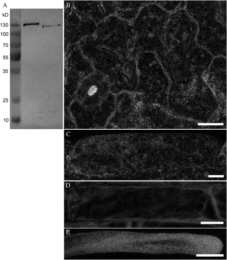
Figure 7.
GFP:VLN3ΔHP, expressed under the control of the VLN3 promoter, shows a cytoplasmic localization. A, Western blotting with an anti-GFP antibody reveals that GFP:VLN3-expressing plants express a fusion protein of approximately 137 kD, the expected mass of GFP:VLN3. GFP:VLN3ΔHP-expressing plants express a fusion protein of approximately 120 kD, which is the expected mass of GFP:VLN3ΔHP. B to E, Representative images of leaf epidermal (B), hypocotyl epidermal (C), and root epidermal (D) cells and a root hair (E) of vln2 vln3 plants in which GFP:VLN3ΔHP is expressed. In these plants, which are not rescued, GFP:VLN3ΔHP fluorescence is equally distributed throughout the cytoplasm. Bars = 10 μm.

