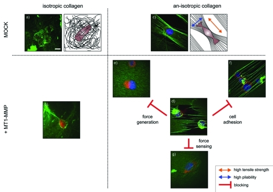
Figure 1.
Representative confocal images and schematic representations of melanoma cells (MV3-clone). The cells express either MT1-MMP (+MT1-MMP) or an empty control vector (MOCK) and were exposed to isotropic or an-isotropic matrices of collagen-I for one hour. An MV3 cell expressing (a) an empty control vector or (b) MT1-MMP on an isotropic collagen matrix show that features of the interaction of the cells with the collagen are difficult to detect. (c) The parallel alignment of collagen fibers causes matrix an-isotropy, with high tensile strength along the fibers and high mobility and pliability of the fibers perpendicular to it (arrows). MV3-cells align along the fibers, but show no features of matrix cleavage under control conditions. (d) Expression of MT1-MMP in cells that are exposed to an an-isotropic matrix induces visible matrix defects. However, pharmacological interference with cell adhesion, force generation or force sensing abolishes the cell’s ability to cleave the fibers. (e) The addition of cytochalasin D breaks down the actin network. The cells are still able to attach to the collagen, but unable to generate any force. (f) Blocking α2β1-integrins with antibodies inhibits the cell’s ability to attach to th collagen. The cells squeeze themselves between the collagen layer and the solid support, a process also known as durotaxis. (g) Blebbistatin inhibits non-muscular myosin. Even low concentrations (1 µM) are enough to interfere with the cell’s ability to cleave and bundle the collagen fibers.(scale bar 10 µm).

