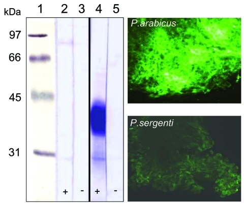
Figure 5.
Left, female sand fly midgut lysates separated by sodium dodecyl sulfate–polyacrylamide gel electrophoresis and blotted onto nitrocellulose membranes. Blots were incubated with biotinylated Helix pomatia agglutinin (HPA) that detects O-glycosylated proteins. Lane 1, molecular mass markers; lanes 2 and 3, Phlebotomus sergenti; lanes 4 and 5, P. arabicus; +, preincubation of lectin with 250 mmol/L N-acetyl-d-galactosamine; –, no preincubation. Right, reaction of fluorescein-conjugated HPA with P. arabicus and P. sergenti midgut cells.

