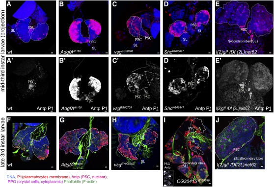
Figure 2 .
Lymph gland phenotypes of Zfrp8null/+ overgrowth suppressors. (A–E, A′–E′) Confocal projections of middle third-instar larval lymph glands. Lymph gland primary lobes are outlined by white dotted line. Normal wild-type glands have few or no plasmatocytes (P1, membrane marker, red A, A′). The number of plasmatocytes in Agdf-Ad11616, vs.gBG00708, and ShcK66847 homozygous mutants (B–D, B′–D′) is drastically increased, compared with that in wild-type (A, A′). PSCs are stained with anti-Antp (nuclear, red). Red channel (Antp and P1) for each projection is shown in (A′–E′). Crystal cells are stained with anti-PPO (cytoplasmic marker, pink). Primary lobes are outlined with white dotted line. Secondary lobes (SL) in (A–D) do not express differentiation markers. (F–J) Confocal cross-sections through the inner layers of late third-instar larval lymph glands. The border between MZ and CZ is shown by yellow dotted line. (F) In wild-type larvae, plasmatocyte maturation is seen in the surface layers (P1, membrane marker, red) of the CZ. Agdf-Ad11616 and vsgEY05552 lymph glands show plasmatocyte differentiation across all CZ layers and also a significant reduction of MZ size (J, H). (I) CG30415EY040039 caused an increase in plasmatocytes, disintegration of the primary lobes, and enlargement and increased differentiation of secondary lobes. The prospective positions of primary lobes (white dotted outline) were identified by the presence of the PSCs (anti-Antp, nuclear red, rectangular inset). (E, E′, J) l(2)gl4/Df(2L)net62 causes lymph gland overgrowth with relatively weak staining for P1 at the surface of the lobes. Staining with phalloidin (green in F–J) shows cell shape allowing detection of lamellocytes and strongly stains heart tube. DNA is shown in blue. Scale bars are 10 μm.

