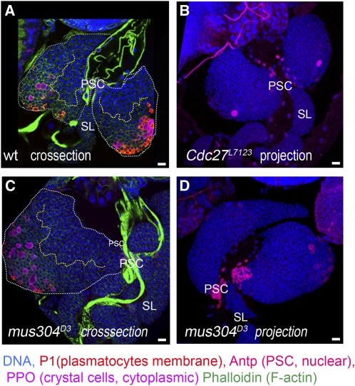
Figure 4 .
Lymph gland differentiation in mus304 and cdc27 mutants Confocal cross-sections (A, C) and projections (B, D) of wild type (A), Cdc27L7123 (B), and mus304D3 (C, D) middle-late third-instar larval lymph glands. Mutant lymph glands show some delay in plasmatocyte differentiation (P1, membrane marker, red). mus304D3 lymph glands show variations in size and differentiation (C, D). PSCs are stained with anti-Antp (nuclear red). DNA is shown in blue. (A, C) The primary lobes are outlined by white dotted line, and the border between MZ and CZ is shown by yellow dotted line; staining with phalloidin (green in A, C) allows detection of lamellocytes. Scale bars are 10 μm.

