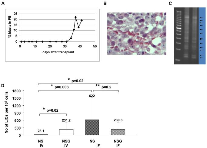
Figure 3.
The G374 line contains high number of LICs in dog (a, b and c) and mice (d). Flow cytometric analysis of blood for GFP+ cells (a) and HE stained BM section (b) of a dog transplanted with YFP-marked (1%) 106 G374 cells. (c) LAM-PCR analysis to track YFP marked LICs in the BM. (d) The number of calculated LICs per 106 G374 cells in NS and NSG transplanted IV or IF. The number of LICs in NSG mice transplanted IV or IF (p=0.02) and NS mice transplanted IF (p=0.003) are greatly increased when compared to the cohort of NS mice transplanted IV. LIC frequency was calculated by Poisson statistics and the method of maximum likelihood using L-Calc software (StemCell Technologies).

