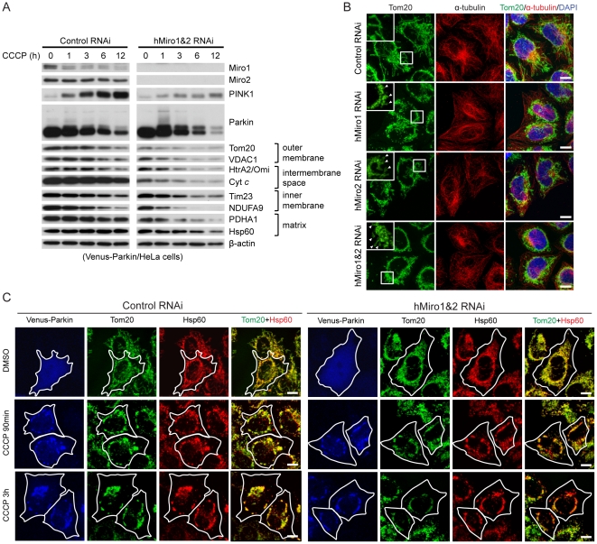
Figure 6. Miro knockdown facilitates the removal of damaged mitochondria by Parkin-mediated mitophagy.
(A) Time course of CCCP treatment to monitor mitophagy in Hela cells stably transfected with Venus-Parkin. Cells were transfected with control siRNA or hMiro1+hMiro2 siRNAs and treated with 10 µM CCCP for the indicated time. Markers for mitochondrial outer membrane (Tom20, VDAC1), intermembrane space (HtrA2/Omi, Cyt c), inner membrane (Tim23, NDUFA9), and matrix (PDHA1, Hsp60) were examined by Western blot to determine the elimination of damaged mitochondria. β-actin was used as a loading control. (B) Immunofluorescence staining showing the formation of perinuclear mitochondrial aggregates after siRNA-mediated knockdown of hMiro1 or hMiro2. HeLa cells transfected with control siRNA, hMiro1 siRNA, hMiro2 siRNA, or hMiro1+hMiro2 siRNAs were stained for Tom20 and α-tubulin. Merged images are shown to the right. Insets show enlarged view of mitochondrial morphology in the boxed areas. Arrowheads indicate ring-like or round-shaped mitochondria. (C) Miro knockdown facilitates the early stage of mitophagy in venus-Parkin transfected HeLa cells treated with CCCP. HeLa cells stably transfected with venus-Parkin were co-transfected with control siRNA or hMiro1+hMiro2 siRNAs and then treated with the DMSO vehicle or CCCP for 90 mins or 3 hrs. Venus-Parkin (blue), Tom20 (green), and Hsp60 (red) were visualized by immunostaining. Merged Tom20/Hsp60 signals are shown to the right. Cells of interest are outlined.

