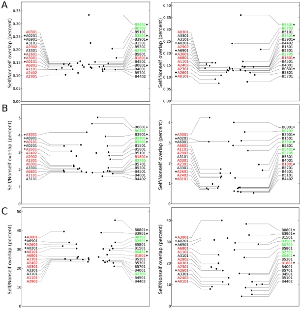
Figure 2. Self/nonself overlaps of peptides presented on different HLA molecules.
In A, the exact overlap of the complete peptide (positions 1–9). In B, the exact overlap of the middle positions of the peptide (positions 3–8) that are assumed to be in contact with the TCR. In C, the degenerate overlap of positions 3–8, i.e. a cross-reactive T-cell overlap. In all cases, the left and right figures show the self/nonself overlaps determined using a scaled or fixed MHC binding threshold, respectively (see Methods). HLA molecules that have been described to have a GC-positive, GC-negative or GC-neutral preference [1] are colored green, red and black, respectively. HLA molecules with additional anchors (see Methods) are indicated with a plus-sign.

