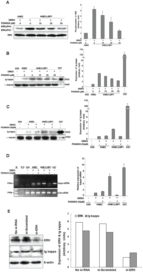
Figure 1. Inhibition of the ERKs signaling pathway blunts LMP1-increased kappa light chain expression at both the mRNA and protein levels.
(A) HNE2-LMP1 cells were treated with the indicated concentrations of PD98059 or 0.1% DMSO for 2 hr. Whole cell lysates were prepared and total and phosphorylated ERKs levels were determined by Western blotting. (B) HNE2-LMP1 cells were treated with the indicated concentrations of PD98059 or 0.1% DMSO for 12 hr. Kappa light chain expression in NPC cells was assessed by Western blotting using a specific antibody. (C) HNE2 and HNE2-LMP1 cells were treated with 50 µM PD98059 or 0.1% DMSO for 12 hr and Western blotting was performed to detect kappa light chain expression. (D) HNE2 and HNE2-LMP1 cells were incubated with medium containing the indicated concentration of PD98059 or 0.1% DMSO for 12 hr. Total RNA was isolated from cells and subjected to RT-PCR, using specific primers designed to amplify kappa light chain and actin mRNAs. (E) HNE2-LMP1 cells were transfected with si-ERK or scrambled oligonucleotide. ERK and Ig kappa protein levels were detected by immunoblotting. The results shown are representative of three independent experiments. Phosphorylation or total expression level for each protein as well as mRNA was estimated by densitometry and are presented as a ratio to the respective loading control (right panels). XG7 and XG6 cells are shown as positive and negative controls, respectively, for kappa light chain.

