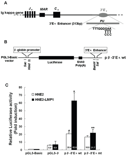
Figure 2. The PU binding site is involved in LMP1-induced human 3′Eκ enhancer activity in NPC cells.
(A) Schematic diagram of the human 3′Eκ core fragment-containing DNA portion used in these experiments. The position of the PU binding site is shown. For simplicity, other protein-binding sites in the 3′Eκ are not shown. The expansion of the PU binding site gives its wild-type sequence and the nucleotides replaced by mutations are underlined. Arrows indicate the nucleotides introduced by mutations. (B) Insertion sites for the DNA fragment in the pGL3-β vector, which contains the human β-globin promoter and the luciferase reporter gene. (C) Comparison of 3′Eκ activity in human nasopharyngeal carcinoma cell lines. The constructs carrying the wild-type PU sequence (pβ-3′Eκwt), mutant PU sequence (pβ-3′Eκmt), pGL3-β or pGL3-Basic with the internal control plasmid pRL-SV40 were transiently co-transfected into HNE2 and HNE2-LMP1 cells. Luciferase reporter assays were performed as described in “Materials and methods”. Values for firefly luciferase activity were normalized to those obtained for Renilla luciferase activity. Values obtained for cells transfected with pβ-3′Eκwt, pβ-3′Eκmt and pGL3-β were divided by the corresponding values obtained for cells transfected with pGL3-Basic. Data are shown as means ± S.D. of three independent experiments performed in triplicate. Statistical significance: #p<0.05 vs. pGL3-β-transfected HNE2 cells; *p<0.01 vs. pGL3-β-transfected HNE2-LMP1 cells; **p<0.05 vs. pβ-3′Eκwt-transfected HNE2-LMP1 cells.

