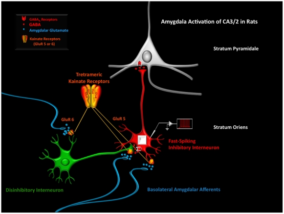
Figure 6. A schematic diagram depicting how an increase of excitatory activity from the BLA might influence the interaction of inhibitory and disinhibitory GABA cells in the SO of CA2/3.
The results reported in this study can be best explained by a model in which BLA afferents influence two types of interneurons: one that is a fast–spiking (FS) inhibitory cell (red) and one that is a disinhibitory neuron (green) that forms GABA-to-GABA interactions with the FS interneuron (1). The diagram suggests that BLA fibers may provide two different KARs-mediated glutamatergic interactions with the disinhibitory neuron. Because PICRO-infused rats showed a significant increase in the amplitude of AHPs in FS-cells, these glutamatergic fibers probably stimulate the KARs located in dendrites of disinhibitory neuron through axodendritic connections (2). Additionally, the further increase in the amplitude of AHPs recorded in FS cells observed in PICRO-treated rats with blockade of the GluR5 subunits of KARs suggests that BLA fibers may also provide a pre-synaptic inhibitory effect of the dysinhibitory axon terminal synapting on the FS cells. This last effect is mediated by GluR5 or 6/7 on GABA-to-GABA terminals (3). BLA fibers have been found to form axo-axonic connections in cortical neuropil (Cunningham et al. 2002) and, in the SO of CA2/3, similar connections with the axon terminations of disinhibitory interneurons maybe present. This circuitry model provides new insights as to how BLA fibers may contribute to the synchronization of oscillatory rhythms generated in the amygdala and hippocampus during normal and abnormal cognitive states.

