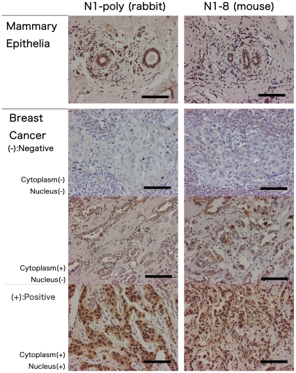
Figure 5. Comparison of immunoreactivities between N1-8 monoclonal and N1 polyclonal anti-RB1CC1 antibodies in formalin-fixed paraffin-embedded breast cancer tissues.
In non-neoplastic mammary ductal epithelium, cytoplasm and nuclei were similarly stained with these two antibodies. In breast cancer tissue samples, these two antibodies reacted similarly in all types of staining variation (I, negative staining in both cytoplasm and nuclei; II, positive staining only in cytoplasm; III, positive staining only in nuclei or in nuclei and cytoplasm). I–II and III were defined as RB1CC1 -negative and -positive, respectively, in the previous clinical cohorts. Bar, 100 µm.

