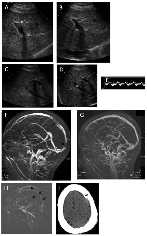
Figure 2.
Selected radiological images. (A) Portal vein thrombosis, patient 2. (B) Portal vein, patient 2, after tPA (C) Middle hepatic vein thrombosis, patient 2. (D) Middle hepatic vein, patient 2, after tPA, with restored flow (E), demonstrated by Doppler. (F) Magnetic resonance (MR) venogram, patient 2. (G) MR venogram, patient 2, after tPA. (H) MR venogram, patient 4, showing extensive venous occlusions. (I) Head computed tomography, patient 4, after tPA, showing a subdural hematoma. Arrows indicate sites of thrombosis in panels A,C, F, and H, and indicate the hematoma in panel I.

