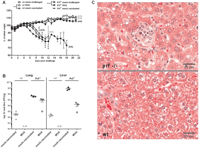
Figure 7. Protective capacity of vaccination is lost in absence of the cytolytic effector molecule perforin.
(A) C57BL/6 mice (wt, open symbols) and perforin deficient C57/BL/6 mice (Prf−/−, closed symbols) were challenged with 3×LD50 ECTV two days after MVA immunization (•), with mock-challenged (▪), and mock-vaccinated (▴) animals as controls. In all experiments weight loss of individual mice was monitored daily (n = 4 to 6 per group). +indicate the individual time of death. Error bars indicate SEMs, and the numbers of surviving/total animals are given in parentheses. Data are representative of two similar experiments. Statistical significance of differences between groups is indicated by * for p-value<0.05, ** for p-value<0.01 and *** for p-value<0.001. (B) At the timepoint of death (day 9 p.i. for Prf−/− mock-vaccinated, day 12 p.i. for Prf−/− MVA and wt mock-vaccinated) or at the end of the experiments (day 21 p.i. for wt MVA) lung and liver were removed, homogenized, and the amount of virus was determined by plaque assay (n = 2 to 3 animals per group). Error bars indicate SEMs and data are representative of at least two independent experiments. n.d.: not detectable. (C) Histopathologic examination of liver tissue from MVA vaccinated and ECTV challenged Prf−/− (upper panel) or wt mice (lower panel) at day 12 p.i.. Tissues were stained with hematoxilin and eosin (HE) and evaluated by light microscopy. Micrographs show representative areas of liver tissue. A typical focus of necrosis and inflammation in the liver of the Prf−/− mouse is visible in the center of the upper micrograph.

