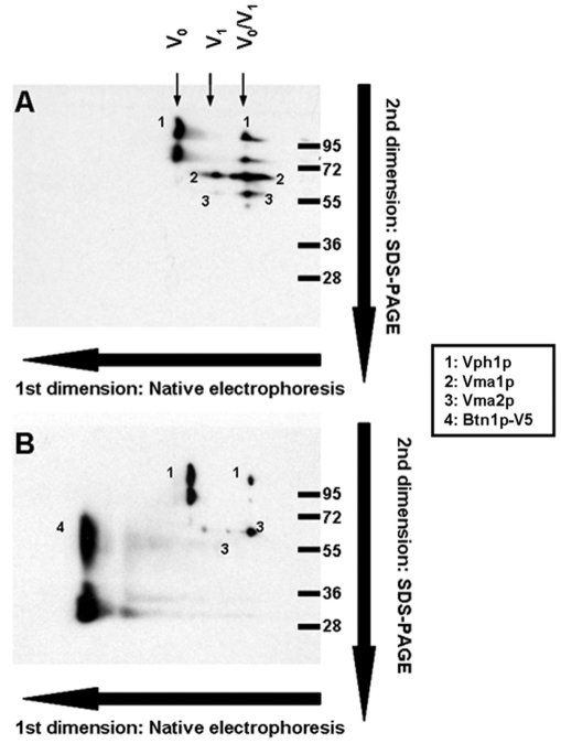
Fig. 1.
Analysis of the protein complexes of the vacuolar proteome by native electrophoresis and second dimension SDS-PAGE. Yeast cells were inoculated at initial A660 0.1 in SC medium and cultured for 24 hours; vacuoles were isolated as indicated in the Methods, and 100 μg samples were analyzed by blue-native electrophoresis to resolve protein complexes followed by a second dimension SDS-PAGE and western blotting using monoclonal antibodies that recognize the V-ATPase subunits Vma1p (69 kDa; V1), Vph1p (100 kDa; V0) and 60-kDa Vma2p (60 kDa; V1), and V5 epitope (46 kDa; Btn1p-V5). (A) Analysis performed in isolated vacuoles from a wild-type strain harboring the empty plasmid pRS426. (B) Vacuoles isolated from a wild-type strain harboring pmBTN1-V5.

