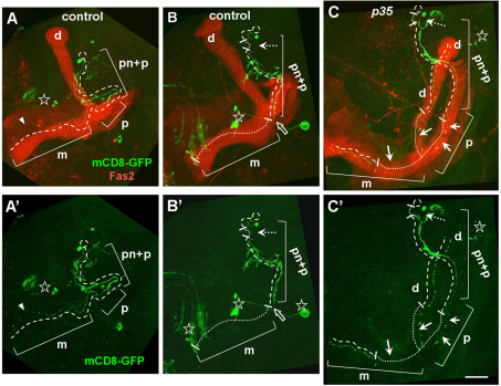
Fig. 5.
Age-related neuronal degeneration. MARCM clones were analyzed in old flies above 80% of mortality of population. Images are Z-stack projections of sequential confocal slices covering the whole MB area. Brain midline is on the left, dorsal side is up. (A,A′) The first sign of degeneration in controls is when the axonal medial (m) lobe of γ-neuron appears thin and partially fragmented (arrowhead). (B,B′) Control γ-neuron with breaks in the proximal neurite (white dashed arrow) and completely missing axonal lobes (white dotted line). Note the end of the existing axon correlated with distal peduncle border (white hollow arrow). (C,C′) p35 mutant MB neuron has multiple breaks in the proximal neurite (dashed arrow), and the dorsal and medial axonal lobes (arrows). Note that the peduncle part of the proximal axon exhibits a reduced amount of fragmentation. Present parts of the proximal neurite (pn), peduncle (p), dorsal (d) and medial (m) axonal lobes are highlighted with a single dashed line. Missing links of neurites are highlighted with fine white dotted line. Other non-MB neuronal types labeled with GFP are marked with a star. Scale bar: 30 μm.

