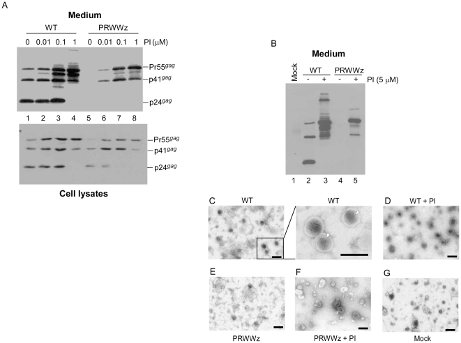
Figure 3. The PRWWz assembly defect is HIV-1 protease activity-dependent.
(A) 293T cells were transfected with wt or mutant plasmids. At 4 h post-transfection, cells were replated on four dish plates and either left untreated (lanes 1 and 5) or treated with the HIV-1 protease inhibitor (PI) Saquinavier at concentrations of 0.01 (lanes 2 and 6), 0.1 (lanes 3 and 7), or 1.0 µM (lanes 4 and 8). At 48–72 h post-transfection, cells and culture supernatant were collected, prepared, and subjected to Western immunoblot analysis. (B–G) Transmission electron microscopy images of concentrated culture supernatant from 293T cells expressing the wt or PRWWz. Culture supernatants from PI-treated or untreated transfectants were collected, filtered, and pelleted through 20% sucrose cushions. Virus-containing pellets resuspended in PBS buffer were subjected to Western immunoblotting (panel B) and transmission electron microscopy. The high-power view (×60,000 magnification) in the inset shows cone-shaped cores (arrowheads) that are characteristic of mature wt virus particles (Panel C). Bars, 200 nm.

