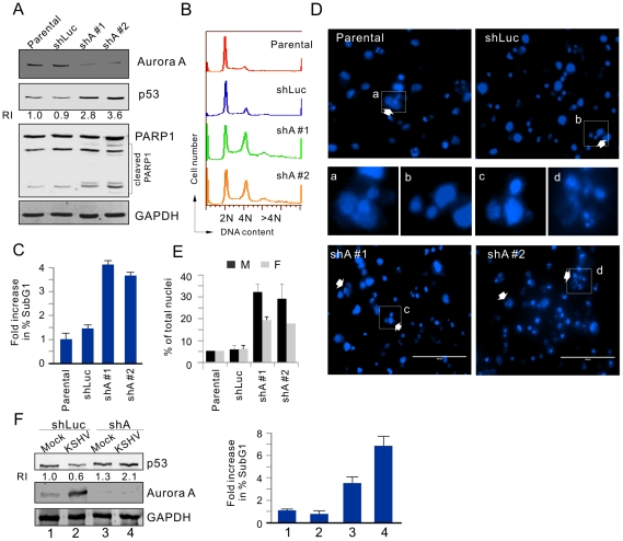
Figure 8. Downregulation of Aurora A induces cell cycle arrest and apoptosis in PEL cells.
(A) Induction of PARP1 cleavage and increased p53 expression in cells with Aurora A knockdown. Parental BC3 cells and BC3 cells transduced by lentivirus expressing small hairpin against Aurora A (shA#1, shA#2) or luciferase (shLuc), were exposed to 0.1% sera for 18 hr. Cell lysates were subjected to immunoblotting as indicated antibodies in the figure. The relative instensity (RI) of p53 protein level is shown. (B) FACS analysis of BC3 cells with or without Aurora knockdown. Aurora A depletion induced cells with greater than 4N DNA content (Propidium iodide staining). (C) Fold changes in the subG1 population of cells with Aurora A knockdown, as analyzed by FACS. (D) DAPI staining of representative fields show multinucleation (white arrows), and inset fields show corresponding photographs to the white rectangles in the upper/lower panels. (E) Quantification of the multinucleated (M) and fragmented nuclei (F) in BC3 cells with or without Aurora A knockdown. (F) Aurora A knockdown dramatically increased p53 accumulation and subG1 population in KSHV-infected not uninfected cells. 293 (Mock) and 293-Bac36 (KSHV) cells were individually transduced by lentivirus expressing small hairpin against Aurora A (shA) and luciferase (shLuc) followed by immunoblotting and subG1 population analysis as described in panels A and C.

