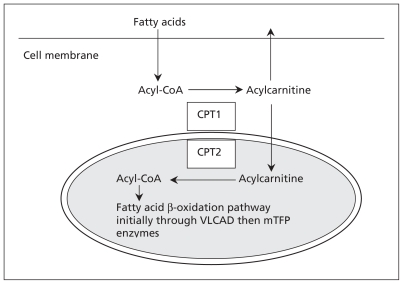
Figure 1:
Simplified illustration of fatty acid β-oxidation. Fatty acids enter cells through specialized receptors. Once in the cytoplasm, fatty acids are activated through esterification (i.e., joined to coenzyme A [CoA] to form acyl-CoA). The acyl-CoA is then linked to a carnitine molecule and this acylcarnitine is shuttled by two enzymes (CPT1 and CPT2) through the outer and inner mitochondrial membranes. The carnitine molecule is removed, leaving the acyl-CoA, which is metabolized by a series of enzymes including very-long-chain acyl-CoA dehydrogenase (VLCAD) and mitochondrial trifunctional protein (mTFP) that consists of hydratase, long-chain 3-hydroxylacyl CoA dehydrogenase (LCHAD) and thiolase activity, into smaller fatty acid chains, which are ultimately converted into adenosine triphosphate (ATP) within the mitochondrion. In our patient, deficiency of LCHAD lead to a backup of long-chain acyl-CoA species that could not be metabolized, and the resultant deficiency of ATP lead to rhabdomyolysis. These long-chain species are converted to acylcarnitine, which is readily permeable to cross the cell membrane and can be detected with a simple blood test. For details about the biochemical pathways of fatty acid β-oxidation, see The Online Metabolic and Molecular Bases of Inherited Disease8 and Online Mendelian Inheritance in Man.9 CPT = carnitine palmitoyltransferase.

