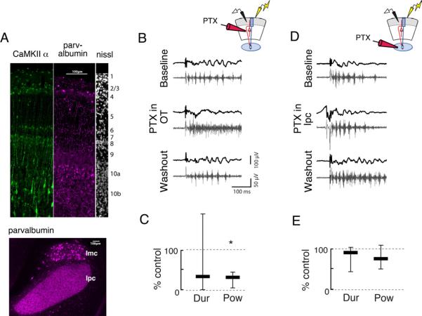
Figure 8. Focal application of picrotoxin to the OT, but not to the Ipc, alters oscillation structure.
Conventions are as in Fig 3.
A) Parvalbumin and CamKII proteins show enhanced immunoreactivity in layer 10 of the i/dOT. (Left, green) CaMKII, a marker for putative excitatory neurons, in the top 10 layers of the OT. (Middle, purple) Parvalbumin, a marker for putative inhibitory neurons, in the top 10 layers of the OT. (Right, white) Nissl stain, showing the layers of the OT. (Bottom) Inhibitory innervation of the Ipc. Parvalbumin is expressed highly in the neuropil in the Ipc, but not in somata. Parvalbumin is expressed somatically in the GABAergic nucleus isthmi pars magnocellularis (Imc).
B) (Left) Gamma oscillations recorded in the sOT of intact slices (Top) are transiently converted to high-frequency events with no appreciable gamma power following a puff of PTX (0.1–1 mM) to the OT (Middle). Gamma oscillations return shortly after the puff (Bottom). (Upper right) Schematic illustrating recording configuration in sOT while puffing PTX in the OT.
C) Duration and power of gamma oscillations is significantly reduced following PTX puff in the OT (duration: 33% of control, power: 31% of control, p<0.001, Friedman test, n=7).
D) (Left) Gamma oscillations recorded in the sOT (top) are not affected by a puff of PTX to the Ipc (middle). (Upper right) Schematic illustrating recording configuration in sOT while puffing PTX in the Ipc.
E) Duration and power of gamma oscillations were unaffected in the evoked response following PTX puff into the Ipc (duration: 88% of control, p>0.3; power: 74% of control, p>0.5, Friedman test, n=7).

