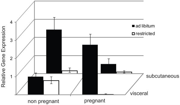
Figure 3. Leptin gene expression in subcutaneous and visceral fat.
At the conclusion of the 10 day diet experiment, subcutaneous and visceral fat were collected and leptin gene expression assessed by real-time RT-PCR. Data were normalized to 18s. All expression is shown as fold-change relative to that in visceral fat of ad libitum fed, non-pregnant mice. Error bars represent 2ΔΔCtCt ±SEM. Comparisons among treatment groups were made by three-way ANOVA and Holm-Sidak post-tests. Diet (restricted vs ad libitum) and reproductive state (pregnant vs non-pregnant) but not fat depot had significant overall effects on LEP expression.

