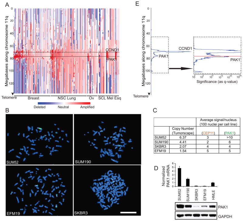
Figure 2. PAK1 is amplified in human cancers.
A. Copy number profile along chromosome 11q of human tumor samples and cancer cell lines that exhibit highest level of PAK1 amplification divided according to cancer type: breast, non-small cell (NSC) lung, ovarian (Ov), small cell lung (SCL), melanoma (Mel) and esophageal squamous (Esq). PAK1 and CCND1 loci are marked. B. FISH of breast cancer cell lines with (SUM52, SUM190) and without (EFM19, SKBR3) PAK1 amplification. Orange fluorescence indicates chromosome 11 centromere reference probe. Green fluorescence indicates PAK1 probe. White bar = 100 μ. C. Tumorscape copy number data is tabulated along with corresponding average number of PAK1 and reference signals from 100 nuclei per cell line. D. mRNA and protein levels of PAK1 in the four breast cancer cell lines and HMLE cells. E. GISTIC analysis of 3131 human cancers along chromosome 11q showing two distinct amplification peaks containing PAK1 or CCND1. The arrow indicates a higher magnification representation of the dotted area to show PAK1 locus (Red).

