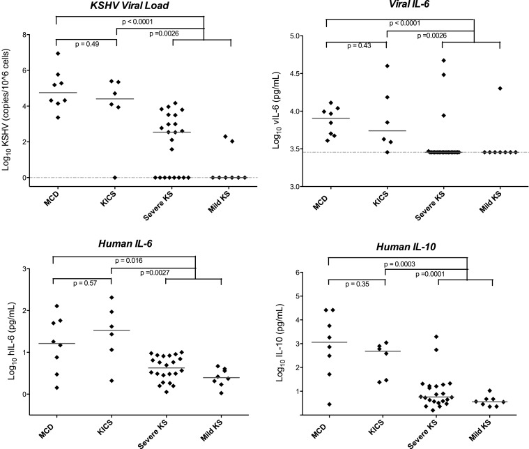
Figure 2.
Viral and cytokine elevations in patients with KICS, KSHV–MCD, and KS. Statistically significant differences in KSHV viral load, viral, and human cytokines were seen in KICS patients compared with controls with KS alone, at levels comparable to those seen in KSHV–MCD. Horizontal lines indicate median value, and the dashed line indicates the lower limit of detection. Values are log transformed; viral and human IL-6 and IL-10 are picograms per milliliter; KSHV VL is copies/106 cells. Note that several of the patients with severe KS and elevated vIL-6 had laboratory abnormalities suggestive of KICS, but did not meet the operative case definition for this series. Modified from Uldrick et al. (2010a), with permission.

